In the CT "super premium" market, the major players focused on different specs and tried to convince hospitals that the future of CT was in this direction (volume/slices/coverage, temporal resolution/cardiac, dose, new imaging/spectral, spatial resolution). Revolution CT is GE's attempt to deliver something that unifies on these goals and they hope to sweep this high end market.
My son died of Bone Cancer when he was 12 years old. For over 4 years I would read and re-read his CT Scans and PET Scans. The issue was his cancer is only curable by surgical removal of any cancer.
The old CT Scan were difficult to interpret and my son's orthopedic doctor (Dr Dorman, CHOP) could look at the scans for 5 seconds and give a answer verified later after surgery. All other doctors (Say around 10) would look over them for 10+ minutes and not be definitive about anything and tell me to go to Dr Dorman at CHOP.
This is big news for the kids and parents of hard tumor cancers. I am really excited that maybe one more kid doesn't have to go through the torture of our current treatment of hard tumors i.e. bone cancer (osteosarcoma) and other horrible diseases.
I always remember the Star Trek film the Search for Spoke when Bones heals the woman in the elevator and yells at the doctors for the barbaric treat of patients. I am thankful for all the doctors we had.
Hi there, great images! I'm curious about how West Kendall Baptist Hospital was chosen for the clinical trial? Is this a single-center study? Lots of angiography in the sample images, so is the trial comparing this to cath?
Great questions. I suspect many operational details were a factor in working with WKBH. Patient load, available space, willing to work with the GE team on issues. CCN [1] in Paris was another trial site. As of now, there's probably a couple dozen installed globally.
Cardiac is one of the big growth areas for CT and Revolution is designed to get better temporal resolution. So absolutely angio imaging is a big part of the publicity. AFAIK there's no direct comparison with cath. But if the Revolution CT cardiac dose was better than the competition (I think it may be), chances are the team will update their dose comparisons between GEHC CT and cardiac cath.
To be fair, while this product is a big improvement, I wouldn't distinguish between it and "traditional CT." It's just the latest evolution of CT (from one particular manufacturer).
Radiation Dose for CT has been a big issue over the past decade. It continues to improve due to better algorithms and hardware changes.
The big features here over GE's other CTs are the fact that it rotates faster and covers more anatomy in a single rotation. Neither of these has a significant impact on the dose. One of the dose oriented features of Revolution is called "ASiR-V" and GE claims that it "is designed to deliver reduced noise levels, improve low-contrast detectability and routinely reduce dose up to 82% for patients of all ages."
CTs are uncommon in family medicine offices. They'd need to forfeit square feet that could otherwise be used for several exam rooms. The offices that do have them probably opt for really low-end ones. So "never"?
As an aside, I think GPs (General Practitioners) are probably phasing out a bit in favor of Physician Assistants and Nurse Practitioners. Though "Family Medicine" still employs lots of MDs.
When said GP has a few million to spend on stuff like this. And when they're no longer needed to send 80% of people home because they have the sniffles and need a doctor to tell them it's just a cold instead of ebola. (Because that's what GP's do in a lot of cases - deal with people who think they've got something worth going to a doctor for).
Yeah, my mom (who is a doctor) was just complaining about this, when she just started out she was a GP in pediatrics and said that 95% of cases were the common cold + over reacting parent, it made it much more difficult to find the interesting cases (read: actually sick kids who needed real treatment), she now works in ICUs and ERs and loves every minute.
This sort of thing is so exciting. I was at the Hunterian (https://www.rcseng.ac.uk/museums/hunterian) last year and it's incredible to see how barbaric fixing (or trying to fix) someone was - and how far it's come in 100 years. Which I hope is much the same as our descendants will be thinking about medicine and surgery now.
That looks very impressive. However, I ask how effective this machine actually is on a medical level. Do these scans provide an advantage over traditional CT scans in that they collect more information? Are they more accurate?
The reason I ask this is because I as a layperson am of course very impressed by the colored, realistic-looking, detailed textured scans -- but will this actually make a difference for a medical/clinical purpose? Will this enable doctors to make better decisions/assessments?
I suspect it will, so I'm very pleased with this innovation. I also note that it might provide more fodder for medical image recognition programs. This tool, combined with good computer vision and analytics, could be very powerful indeed.
The article is a little light on detail. However I see a cardiologist every couple of years due to a strong family history of isochaemic heart disease. He's been putting off more invasive scans since nothing in my medical presentation suggests I actually have a problem, but he said that this sort of scan where they'll be able to look for blockages on moving arteries will be a game changer for cardiac diagnosis - it's the very fast scanning on moving tissue that will be the big improvement. It's not clear whether this new machine actually provides that from the article, but he was expecting it to be developed and in use within the next couple of years.
GE claims 0.23mm spacial resolution across a 160mm span in 0.28 seconds if I understand correctly [1]. This would be about 695^3 voxels. Seeing the example captures [2] I guess the device can scan multiple volumes in a row to cover larger spans.
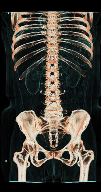
Yes I agree very nice images. The article was light on detail, but looking at the results it appears, in common with other x-ray imaging, to be most effective with dense tissue, like bone where it reveals fine detail.
Soft tissue isn't as well delineated. No doubt that the vascular images were made using contrast media. The value of the new device may well be most evident in the images of vessels in the brain, a region notoriously difficult to visualize adequately. Those images are things of beauty and I can see how the technology could translate to saved lives. (Think about enhanced ability to find a brain aneurism or small bleed.)
MRI is complementary as more effective with soft tissue than x-ray, but less useful for dense matter like bone. Amazing how much the world of medicine has changed in the last 40 years. It's a domain where digital technologies have made a startling difference. Great to see these wonderful technologies put to such good use.
Yep. I've often fantasized about a day when imaging and scanning tech is at the point where you can obtain a high resolution, three dimensional scan of a person's body and then apply software analysis that can detect all sorts of potential issues.
Sure, I basically described Star Trek or any number of sci-fi med bays but when comparing the sorts of internal imaging we could do 50 years ago to what can be done today, it makes me wonder what might be possible 50 years from now. Definitely just daydreaming on my part but seeing things like modern medical imaging always makes me think of it.
Correct me if I'm wrong, but this just looks like a modern CT scan. Impressive, of course, but definitely not a revolution. These machines have been steadily improving for thirty years.
You are absolutely correct. Revolution is strictly a brand name. 160mm and 0.28s is an incremental improvement over GE's CT750HD, albeit in two dimensions.
CT scan time is ~0.2-0.5 seconds for a 160mm slice, so a whole body can probably be scanned fairly quickly. However, CT scanning is only done for the body parts of interest, to keep radiation exposure low.
Most fascinating here is the ability to capture a beating heart!
It's also computationally intensive to reconstruct all the x-ray images into a 3D model. I worked for a company that wrote software to speed this up with GPGPUs. Standard software packages took on the order of hours for one scan, while parallelism brought it down to 10s of minutes - this was just to transform the x-ray data into voxels. Not sure how long the post-processing to identify & highlight organs would take.
It doesn't matter so much for humans, though. The main market for the above-mentioned software product was research labs imaging hundreds of lab rats.
The pretty pictures in the article can be rendered in real time using GPUs. Unless the pictures here use techniques that I haven't heard about, it is done by treating the voxels as a semi-transparent cloud of different-colored materials and using a fragment/pixel shader to simulate rays of light projected through this volume. It's pretty straightforward; the volume data is stored in a 3D texture and each ray of light is simulated using one "pixel" of the fragment shader.
This is enhanced by assigning more weight to the voxels that are in a boundary region between tissues of different density (you see this effect in the pictures of lungs, kidneys etc; the organ is transparent but the edges are visible) and applying a shading model to these boundary regions to improve perception of their spatial orientation.
Different organs can be pre-selected by marking their constituent voxels, and different models/colors/shading applied to each organ. This process can also sometimes be automated, if you have the luxury of a table that correlates voxel intensity (x-ray absorption value) with tissue type (bone, muscle, air, etc).
It's much, much simpler than that. You just project a ray from the X-ray point source through the center of a voxel onto the detector, interpolate the X-ray energy detected, and add that energy to the sum in that voxel. Repeat for each voxel and each x-Ray taken (CT scans take a bunch of x-Rays going around in a circle), remove some noise, and boom you've voxelled the data. Fundamentally you're just summing the energy that passes through each point in space. It's trivially parallelizeable.
Yes, my comment only treats the process from the point where you have a complete dataset of voxels to the point where you have a finished rendering. ahelwer didn't treat this part of the process in his comment.
Unfortunately, there's a tradeoff: each imaging session exposes the patient to radiation. Too much radiation can be problematic.
The article does mention lower exposure but it's not clear whether it's just shorter in time (and more intense) or is the total level of radiation actually lower.
The dose is primarily a function of the power delivered to the X-Ray source, the size of the beam and the scan's duration. The big features of this device are coverage (160mm) and rotation speed (mostly dose neutral). If you were imaging 160mm before you had to use multiple sequential scans, so the dose is not significantly different from that scenario.
Revolution has similar or superior (meaning less) dose than GE's previous high end scanner. GE's high end scanners tend to offer better dose than the competition.
IIRC on this scanner, the actual data collection is very fast (few minutes) but the high-quality image reconstruction needs just under an hour. The images in the link are also heavily manually post-processed which is labor intensive.
GE sells a low-dose feature branded "Veo" that has ~30-minute reconstruction times. CT750HD spectral imaging (branded GSI) takes as long as 20-minutes.
Revolution imaging takes on the order of minutes (2-3). Indeed the 3d post-processing takes time and probably includes many manual steps/tweaks.
Hmm, that's too bad. I could've sworn it had hit the 30-minute mark. There haven't been many refreshes so I don't know that it's been improved since the initial release.
Well, IMO, after five or so minutes there's not a huge difference between 30 and 60. But it does depend a bit on how the imaging workflow goes (do you wait for those images before letting the patient leave?, e.g.).
But you can get some pretty dramatic image quality or dose improvements from Veo, so maybe it's worth it.
In the US I think that they may be by prescription-only? Maybe not, worth a call to your local outpatient diagnostic imaging center. Don't call a hospital, they'll charge you too much.
I'd guess you could get one from any-old-CT-scanner for something like $500 to $1000? Someone who just bought this Revolution scanner will probably charge much more. Apparently some people get charged upwards of 10k USD [1]!
But honestly if you just want images of your guts you'd be fine with any old CT.
Once you have the 2d axial images, you can probably find open source software that will do the 3d projection.
You probably don't want to get one without a good reason: this uses x-rays, and you get a considerably-higher dose of radiation than something like a normal chest x-ray. As for other comments saying an older machine would be cheaper, etc., this is true, but older machines actually deliver more radiation.
All that being said, you can get modern CT scans cheaply in countries other than the US. In Japan, for example, there are ex-pat clinics that give comprehensive medical checkups, which include chest CT scans, starting around US$650, which is absolutely incredible compared to costs here in the States.
later edit: dear downvoters, you only get gray levels from a CT and those pretty colors assigned after what I assume is automated segmentation don't improve the diagnosis. I'd guess that any segmentation errors make the visualization more difficult.
The colours provide contrast. It makes it much easier for a doctor to find the parts they are interested in as quickly as possible. Not to mention the fact that doctors aren't always interested in the tiniest imperfections and may be looking for large changes.
When you have things segemented out and coloured, it is also much easier to view the object, be it an organ, blood vessel, etc. in 3 dimensions, which is something that is very useful.
Admittedly, they can get in the way sometimes, but that's why it is an optional feature.
In the CT "super premium" market, the major players focused on different specs and tried to convince hospitals that the future of CT was in this direction (volume/slices/coverage, temporal resolution/cardiac, dose, new imaging/spectral, spatial resolution). Revolution CT is GE's attempt to deliver something that unifies on these goals and they hope to sweep this high end market.