That trichinella cyst photo [1] is by Nathan Myhrvold [2], who is a name some here might recognize. He got rich as an early executive at Microsoft, used that to start a patent troll firm, and then got really into cooking.
His six-volume "Modernist Cuisine" is like the "The Art of Computer Programming" for molecular gastronomy.
Travelling the universe at this scale seems as important and amazing to me as travelling to a distant star. The igniting matchstick image has so much going on!
One cringe though ... the images all seem excessively post-processed to me. Unrealistically saturated, contrasted, sharpened, etc. The colour, especially, just seems over the top and I feel like seeing these microscopic subjects enlarged is mind-blowing enough without the postprocessing distraction which makes it seem less real.
One thing to note is that the fluorescence images like https://www.nikonsmallworld.com/galleries/2023-photomicrogra... are false color. In those systems, you typically excite a specific fluorophore, one at a time (once for each type in the sample), and read out the result with a monochromatic camera through frequency-specific filters. The final image is composited with the grayscale intensity mapped to color value.
I don't think the microscopy images underwent sharpening, although I'm not certain.
My friend who works in the field prefers to look at individual color planes in monochrome (no color at all) rather than the super-saturated composites.
They can be somewhat true color if you reconstruct the image with the appropriate human perceived color for the wavelength the dye emits, doesn't work well for multiple dyes with close wavelengths.
I'm of two minds about the processing... if adding lots of artificial color, sharpening, and contrast brings out the detail(s) the researcher is looking for without actually altering them, then it seems like fair game.
But yes, the raw photos would be nice to see as well, just for comparison's sake. That may be easier said than done given the prevalence of image stacking.
To my untrained eye I don't understand why #1 is #1, #2 has more interesting details and demonstrates much more skills. Perhaps I don't understand the evaluation parameters though, #1 is probably a bigger technical achievement.
Not sure it demonstrates much more skills (lighting a match vs. extracting an optical nerve and differentiating it to proteins, glials and vasculature).
Note that the list of techniques used for the matchstick includes "image stacking", which I thought involves multiple exposures, but a burning match is not quite a static subject that would remain constant across multiple shots. I am curious what setup they have done to pull it off.
Related, this photographer has videos of macro shots of burning match elsewhere:
The 16th place, "Carbon Nanotubes" really struck me as being generated by some AI. It has many of these hallmark "hallucination"-like features that we find in AI images.
Am I not able to distinguish between reality and AI anymore?
Not taking away anything from human ability to create beautiful art but nature is literally light years ahead. Spent yesterday in an average aquarium and was blown away by jellyfish alone.These guys, too. https://share.icloud.com/photos/018gc-y5yDp7TMEaTTuCL9Kog
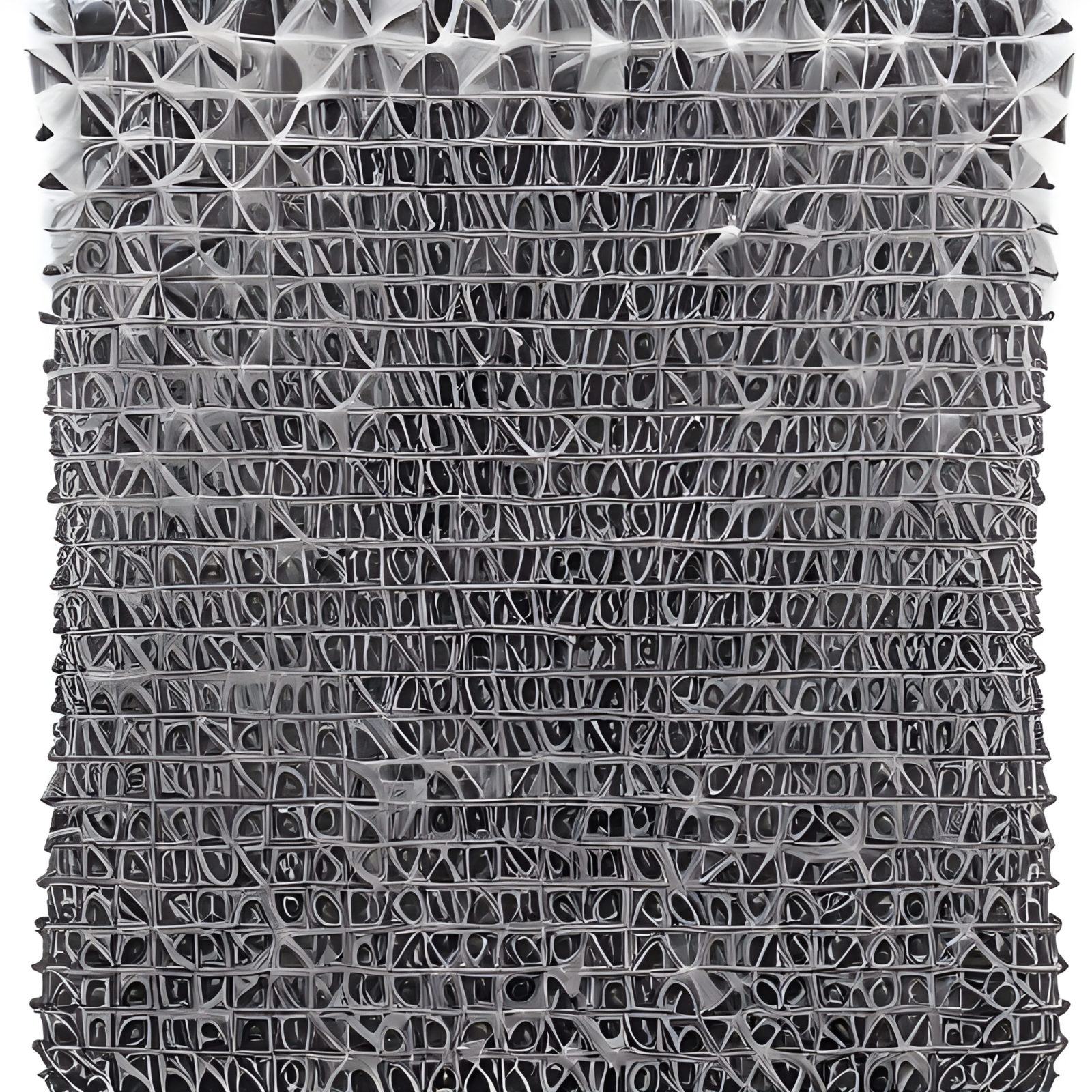
Anyone with chemistry chops here able to explain what I'm seeing in #11 "Crystallized sugar syrup"? I do macrophotography myself and know roughly what 25x means, but I'm having trouble even parsing the scale of these structures. Sugar crystallizes in sheets?
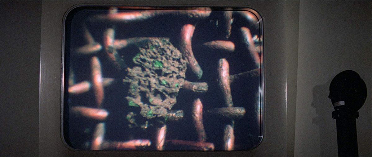
I wondered the same thing but then I concluded it was coincidental. If you're trying to filter for meteorites, you're going to be using that exactly hardware (a wire mesh basket), using a level of magnification that gives a view of the retained object, whose size is similar to the mesh screen hole diameter.
I would imagine that the props department went to a nearby micrometerologist who gave them a basket with a micron mesh and maybe some small volcanic rocks.
As an amateur photographer (mostly black and white film of my kids, dogs, and family) I'm struck by how incredible these look. The lighting, the brightness of the images, clarity, everything... it's beyond what I thought was possible through a microscope.
If there is anyone here who takes images like this, could you describe what goes into getting lighting and photographs like this?
what would be the cheapest way to get into this as a hobby ?
I understand hobbies are expensive but sometimes with minimal time, you wanna reduce costs.
Start with a clip-on macro lens for your smartphone. See if you find it fun to actually frame and adjust your shots, and to patiently find good subjects. If you don't enjoy those parts, I wouldn't bother getting a digital camera. You'll want to spend well over $1k on a dedicated setup if you're going for macro photography and you already have a smartphone, so experiment before diving in!
I did phone-only for about 2 years before getting a nice lens and decent camera (the lens is the important part). I wouldn't say you need to spend that long, but if it doesn't become a hobby without a dedicated camera, it won't become a hobby with one.
His six-volume "Modernist Cuisine" is like the "The Art of Computer Programming" for molecular gastronomy.
[1]: https://www.nikonsmallworld.com/galleries/2023-photomicrogra...
[2]: https://en.wikipedia.org/wiki/Nathan_Myhrvold